Resources
Stay informed
Dix-Hallpike Maneuver
Contents
Test protocol
Context
Benign paroxysmal positional vertigo (BPPV) is the most common of all vertigo conditions: it is a true rotational vertigo, triggered by changes in head position, lasting less than 60 seconds, associated with nystagmus, without cochlear or neurological signs.
According to the HAS (French national authority for health), it disappears spontaneously in 30% of cases within seven days.
It involves one or more semicircular canals and causes different types of associated symptoms, such as nausea or vomiting.
Physicians and physical therapists must refer any patient presenting with BPPV to a specialized professional if they have not received specific training.
BPPV may be caused by otolithic debris in the semicircular canal of the ear that continues to move after the head has stopped moving. This results in ongoing motion that conflicts with other sensory inputs, particularly visual and somatosensory information.
The semicircular canals are filled with a fluid called “endolymph.” The main sensory organ of each canal is called the “ampullary crest,” which is stimulated by the movement of the cupula.
Head movement causes relative movement of the endolymph within the semicircular canal, which bends the cupula and the hair cells embedded in the ampullary crest and causes depolarization or hyperpolarization of the corresponding vestibular nerve. This is how we receive information about the position of our head in space.
Whether idiopathic, post-traumatic, or infectious, BPPV is thought to be caused by the displacement of otoliths (natural components composed of calcium carbonate and present on the otolithic maculae) into the semicircular canals, particularly the posterior canal. This is referred to as “canalolithiasis.” More rarely, these otolithic debris may adhere to the cupula, in which case it is referred to as “cupulolithiasis.”
In canalolithiasis, it is assumed that freely floating debris within the semicircular canal acts like a piston, causing continued movement of the endolymph even after motion has stopped.
This results in the movement of the cupula and deflection of the hair cells, thereby causing vertigo.
Video of the Test
Execution
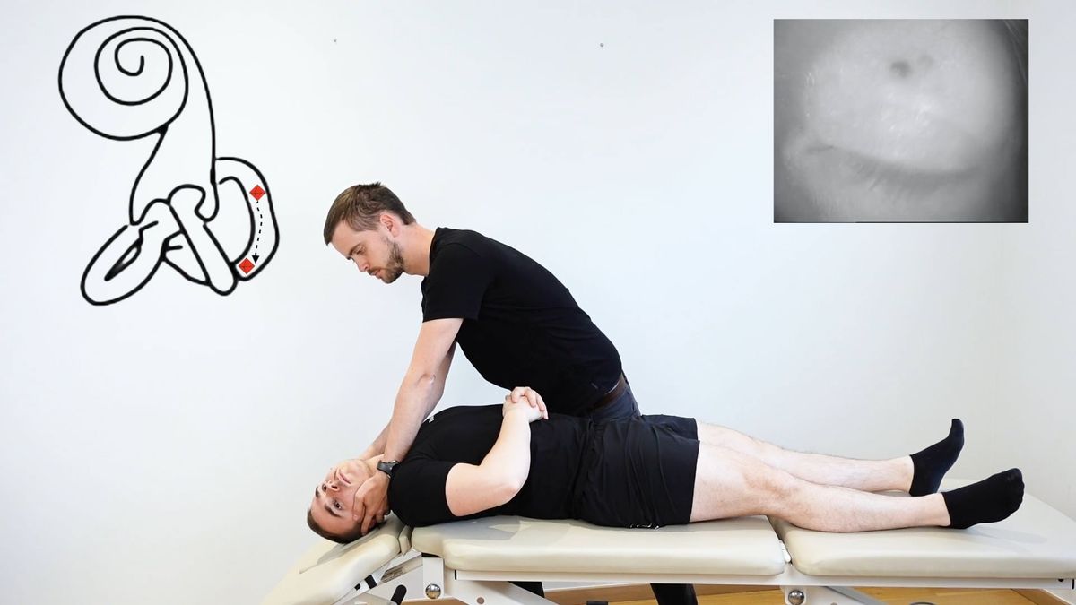
To perform this maneuver, the patient is seated on a table with the headrest lowered a few degrees below horizontal.
To perform a right Dix-Hallpike, the head is rotated 45° to the right, and the patient is laid in supine while maintaining the 45° right head rotation. At the end of the movement, the patient’s head rests in extension on the table’s headrest. By turning the head 45° to the right, the right posterior semicircular canal and the left anterior semicircular canal (i.e., the LARP plane) are aligned with the sagittal plane, which is the plane of movement in the Dix-Hallpike Maneuver.
Similarly, for a left Dix-Hallpike, the head is rotated 45° to the left, and the patient is laid supine while maintaining the 45° left head rotation. In this way, the right anterior semicircular canal and the left posterior semicircular canal are positioned in the plane of movement.
We wait approximately 20 seconds to observe whether nystagmus or vertigo appears. If so, we analyze the direction of the nystagmus; otherwise, the patient is brought back up to a seated position.
For a therapist who does not have a table with a declining headrest, the maneuver is performed by allowing the patient’s head to hang over the edge of the table once in supine. In this case, the maneuver is less comfortable for the patient and offers less freedom for the therapist, who will often need to support the patient’s head.
Clinical Value
The Dix-Hallpike Maneuver is considered a reference test for the diagnosis of BPPV of the posterior semicircular canals (Evren et al., 2017; Talmud et al., 2022). In the literature, this test has been associated with a sensitivity of 82% and a specificity of 71% in the diagnosis of posterior canal BPPV, as well as a positive predictive value of 83% and a negative predictive value of 52% (Hanley K., O’Dowd, 2002; Lopez-Escamez et al., 2005; Evren et al., 2017). Its low NPV indicates, however, that a negative result on the Dix-Hallpike Maneuver does not rule out a diagnosis of posterior canal BPPV (Evren et al., 2017).
Bibliography
Evren, C., Demirbilek, N., Elbistanlı, M. S., Köktürk, F., & Çelik, M. (2017). Diagnostic value of repeated Dix-Hallpike and roll maneuvers in benign paroxysmal positional vertigo. Brazilian Journal of Otorhinolaryngology, 83(3), 243‑248.
Lopez-Escamez J.A., Gamiz M.J., Fernandez-Perez A., Gomez-Finana M. Long-term outcome and health related quality of life in benign paroxysmal positional vertigo. Eur Arch Otorhinolaryngol. 2005;262:507–511.
HAS. Vertiges positionnels paroxystiques bénins : Manœuvres diagnostiques et thérapeutiques. Argumentaire. Décembre 2017
Hanley K., O’Dowd T. Symptoms of vertigo in general practice: a prospective study of diagnosis. Br J Gen Pract. 2002;52:809–812.
Halker RB, Barrs DM, Wellik KE, Wingerchuk DM, Demaerschalk BM. Establishing a diagnosis of benign paroxysmal positional vertigo through the Dix-Hallpike and side-lying maneuvers. A critically appraised topic. Neurologist 2008;14(3):201-4.
Helminski JO, Zee DS, Janssen I, Hain TC. Effectiveness of particle repositioning maneuvers in the treatment of benign paroxysmal positional vertigo: a systematic review. Phys Ther 2010;90(5):663-78.
Lopez-Escamez J.A., Gamiz M.J., Fernandez-Perez A., Gomez-Finana M. Long-term outcome and health related quality of life in benign paroxysmal positional vertigo. Eur Arch Otorhinolaryngol. 2005;262:507–511.
Ibekwe TS, Rogers C. Clinical evaluation of posterior canal benign paroxysmal positional vertigo. Niger Med J 2012;53(2):94-101
Talmud, Jonathan D., Ryan Coffey, et Peter F. Edemekong. « Dix Hallpike Maneuver ». In *StatPearls*. Treasure Island (FL): StatPearls Publishing, 2022.