Resources
Stay informed
FABER Test
Contents
Test protocol
Context
The letters of the FABER Test stand for “flexion,” “abduction,” and “external rotation.” When associated with other tests, this combination of three passive movements produces a pain provocation test that is used to assist in the diagnosis of hip conditions, such as involvement of the coxofemoral joint, lumbar region conditions, and sacroiliac joint conditions.
Video of the Test

Execution
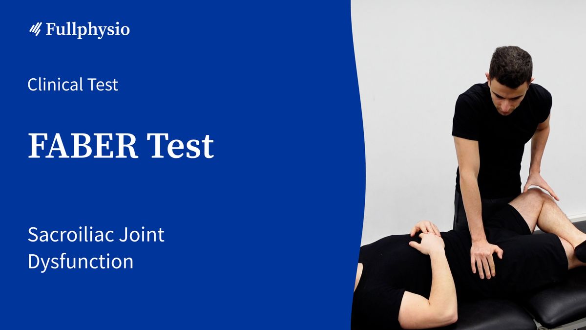
To perform this test, the patient is in supine. The leg is placed in a figure-4 position, with the hip flexed and abducted, and the ankle of the tested leg resting on the contralateral thigh, just above the knee.
The physical therapist places their cranial hand on the opposite anterior superior iliac spine (ASIS) to stabilize the pelvis, then gently applies downward pressure on the knee of the tested limb toward the table until full range of motion is reached.
In this position, the physical therapist can add a few additional small-range oscillations to assess pain provocation at end range.
The test is positive if it reproduces the patient’s symptoms or if the range of motion is limited. This may indicate an intra-articular hip problem, a sacroiliac joint problem, or an iliopsoas contracture.
Clinical Value
In 2020, Nejati and colleagues sought to evaluate the reliability and validity of palpation tests during movement, such as the Gillet Test and sacroiliac joint provocation tests, using anesthetic blocks of this joint as the reference standard.
This study showed that the FABER Test had the highest specificity and positive predictive values of all the provocation tests examined. Furthermore, a combination of the FABER Test and the Thigh Thrust Test improved overall diagnostic accuracy.
The authors emphasize that a greater number of positive physical tests contributes more to the diagnosis of sacroiliac joint condition. It is also recommended to use at least 3 provocation tests in addition to palpation tests during movement to confirm a sacroiliac joint diagnosis. Finally, as the existing literature reports inconclusive findings for the use of individual physical tests, it is advised to use a combination of these tests in conjunction with other data sources, including patient interview, symptom description, and imaging, to diagnose sacroiliac joint dysfunction.
Bibliography
Dreyfuss, P., M. Michaelsen, K. Pauza, J. McLarty, and N. Bogduk. “The Value of Medical History and Physical Examination in Diagnosing Sacroiliac Joint Pain.” *Spine* 21, no. 22 (November 15, 1996): 2594–2602.
Martin, Robroy L., and Jon K. Sekiya. “The Interrater Reliability of 4 Clinical Tests Used to Assess Individuals with Musculoskeletal Hip Pain.” *The Journal of Orthopaedic and Sports Physical Therapy* 38, no. 2 (February 2008): 71–77.
Maslowski, Erin, William Sullivan, Jeri Forster Harwood, Peter Gonzalez, Marla Kaufman, Armando Vidal, and Venu Akuthota. “The Diagnostic Validity of Hip Provocation Maneuvers to Detect Intra-Articular Hip Pathology.” *PM & R: The Journal of Injury, Function, and Rehabilitation* 2, no. 3 (March 2010): 174–81.
Nejati, Parisa, Elham Sartaj, Farnad Imani, Reza Moeineddin, Lida Nejati, et Marta Safavi. « Accuracy of the Diagnostic Tests of Sacroiliac Joint Dysfunction ». Journal of Chiropractic Medicine 19, nᵒ 1 (mars 2020): 28‑37.
Telli, Hilal, Serkan Telli, and Murat Topal. “The Validity and Reliability of Provocation Tests in the Diagnosis of Sacroiliac Joint Dysfunction.” *Pain Physician* 21, no. 4 (2018): E367–76.
Dutton M. Orthopedic: Examen, évaluation et intervention. 2e éd. New York: The McGraw-Hill Companies, Inc; 2008.
Flynn T, Cleland J, Whitman J. User's guide to the musculoskeletal examination: Fundamentals for the evidence-based clinician. Buckner, Kentucky: Evidence in Motion; 2008
Mitchell, Bruce, Paul McCrory, Peter Brukner, John O’Donnell, Emma Colson, and Robert Howells. “Hip Joint Pathology: Clinical Presentation and Correlation between Magnetic Resonance Arthrography, Ultrasound, and Arthroscopic Findings in 25 Consecutive Cases.” *Clinical Journal of Sport Medicine: Official Journal of the Canadian Academy of Sport Medicine* 13, no. 3 (May 2003): 152–56.