Resources
Stay informed
Heel-Contralateral Knee Maneuver (HCLK)
Contents
Test protocol
Context
Piriformis syndrome is considered the equivalent of an entrapment neuropathy involving compression of the sciatic nerve in the infrapiriform foramen. It presents as sciatic-type pain originating in the gluteal region.
This condition remains poorly understood from an anatomical, biomechanical, and clinical standpoint, and its diagnosis is a subject of controversy.
Multiple causes of sciatica may coexist, each contributing significantly to the patient's symptoms. When examining the potential causes of sciatic nerve compression, it becomes apparent that many structures may be involved, not just the piriformis. Recent publications suggest that the term “deep gluteal syndrome” may better describe this constellation of muscle- and nerve-mediated pain (Martin et al., 2015). Therefore, piriformis syndrome would be a subgroup of deep gluteal syndrome (Hernando et al., 2015).
Deep gluteal syndrome is characterized by pain or dysesthesia in the glutes, hip, or posterior thigh, or radicular pain due to non-discogenic sciatic nerve compression in the subgluteal space (Hernando et al., 2015). These symptoms fluctuate throughout the day and vary with changes in position (Michel et al., 2013a).
In their 2013 study, Michel et al. developed the idea that the piriformis muscle, initially a lateral hip rotator, becomes an internal rotator beyond 90° of hip flexion and would, therefore, be stretched by a position of lateral rotation. Based on this observation, they created the Contralateral Heel-to-Knee Maneuver in an attempt to compress the sciatic nerve against the piriformis muscle (Michel et al., 2013b).
Before performing this test, it is recommended to examine and rule out the most common conditions of the lumbar spine and sacroiliac joint that could explain the patient’s symptoms.
Video of the Test

Execution
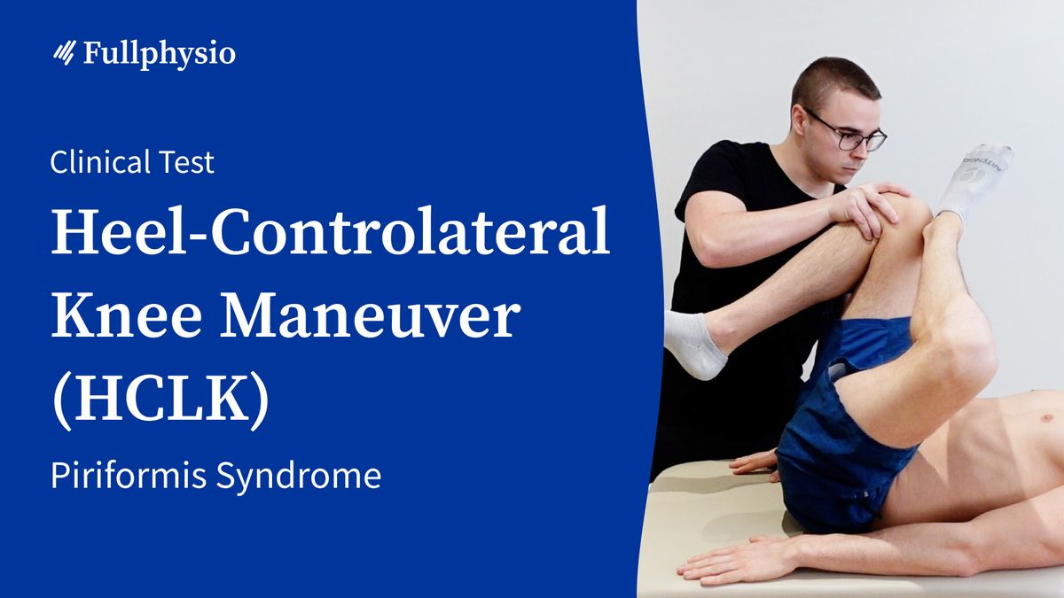
To perform the test, the patient is in a supine position with the knees flexed. They perform external hip rotation with the affected limb and place their heel on the contralateral knee. The physical therapist then flexes the contralateral hip to reach the maximum range of motion.
The position must be held for several tens of seconds to trigger the characteristic gluteal pain and sciatic radiation (Michel et al., 2013a, 2013b).
The test is positive when it reproduces the patient’s symptoms (Michel et al., 2013b).
Clinical Value
In their 2013 study, Michel et al. associated this test with a sensitivity of 100% and a specificity of 60% for diagnosing piriformis syndrome in a symptomatic population (Michel et al., 2013a).
However, this test does not allow the piriformis muscle to be isolated from the other short external hip rotators. The nerve could, therefore, be compressed into a narrow muscle passage located between the inferior border of the piriformis muscle and the obturator internus, gemelli, and quadratus femoris muscles, around which it wraps.
Based on this observation, Michel et al. recommend using this test in combination with other tests, such as the FAIR Test or the Feisberg Test, which stretch the piriformis in internal hip rotation (Michel et al., 2013a).
Bibliography
Hernando, Moisés Fernández, Luis Cerezal, Luis Pérez-Carro, Faustino Abascal, et Ana Canga. « Deep Gluteal Syndrome: Anatomy, Imaging, and Management of Sciatic Nerve Entrapments in the Subgluteal Space ». Skeletal Radiology 44, no 7 (juillet 2015): 919‑34.
Martin, Hal David, Manoj Reddy, et Juan Gómez-Hoyos. « Deep Gluteal Syndrome ». Journal of Hip Preservation Surgery 2, no 2 (juillet 2015): 99‑107.
(a)Michel, F., Decavel, P., Toussirot, E., Tatu, L., Aleton, E., Monnier, G., … Parratte, B. (2013). Piriformis muscle syndrome: Diagnostic criteria and treatment of a monocentric series of 250 patients. Annals of Physical and Rehabilitation Medicine, 56(5), 371–383.
(b)Michel, F., Decavel, P., Toussirot, E., Tatu, L., Aleton, E., Monnier, G., … Parratte, B. (2013). The piriformis muscle syndrome: An exploration of anatomical context, pathophysiological hypotheses and diagnostic criteria. Annals of Physical and Rehabilitation Medicine, 56(4), 300–311.